Working hours

9-6 PM Monday to Saturday

Periapical x-ray

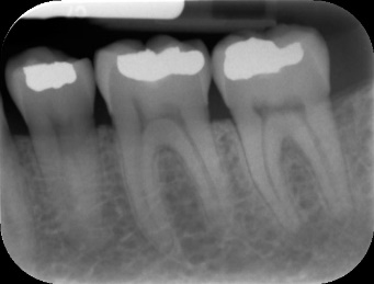
Periapical x-ray is a type of x-ray that shows one to two teeth from their crown to their root and the surrounding bone. It is a very commonly used diagnostic measure. This type of x-ray can be used for any tooth in the mouth and it is mostly used to determine the depth of the decay and if the tooth needs endodontic therapy, if there are any periapical changes, impacted teeth, cysts and more.

This is another very important diagnostic X-ray in dentistry, that dentists use on daily basis. Compared to the panoramic X-ray, the periapical shows only two, up to three teeth. And it presents a very detailed picture of all the hard tissues. It comes from the Greek word “peri” which means around and “apex” which means tip.

Uses:

The main use is to diagnose conditions of teeth and the surrounding tissues. Sometimes dentists are not sure whether there is a periapical process, so they have to use this X-ray to set up a diagnosis. Other times, they use it as a part of the treatment, for example, when they need to see dental caries and its depth. When it comes to decay, dentists can monitor which tissues it has affected, which surfaces, and whether there is some proximity to the dental pulp. This is all very useful when removing the decay and placing the filling. For periapical conditions, an x-ray can show if the infection has spread from the pulp to the surrounding bone, whether it is a localized infection or it has spread. It helps with diagnosing fractures of the teeth and the bone, cysts, tumors, the number of roots, number of canals, the location of the apex and more.

Read more

periapical-x-ray

Procedure:

The procedure is very simple. This is an intraoral type of X-ray, where the film is inside the mouth. The X-ray technician will give you all the instruction, place the film inside the mouth and adjust the tube. You have to take off all metal things you have on you before the procedure including jewelry.